High-speed imaging – QRI –
QRI (Quick Raman Imaging) utilizes the combined technology of high-speed and high-accuracy stage and high-speed data acquisition CCD detector to obtain imaging data of more than tens of thousands measurement positions in just a few minutes. This method is useful for a wide range of samples with sizes from millimeters to sub-microns.
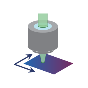
Faster imaging over wide area
The QRI reduces measurement time significantly for wide range of sample imaging measurement.
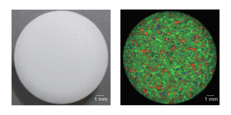
Pharmaceutical tablet (left: observation image, right: chemical image)
(red: acetaminophen, green: etenzamide, blue: caffeine)
Details in narrow area
When analyzing a sample microstructure, QRI’s ability to perform the sample measurements at small intervals and high-speed scanning of the sample stage enables the acquisition of high-spatial resolution and high-resolution images in a short amount of time.
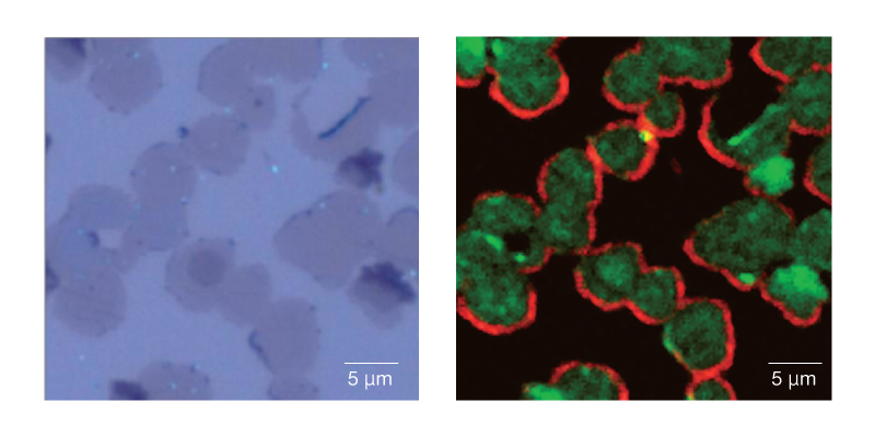
Graphene (left: observation image, right: chemical image)
(red: D band, green: G band)
Laser scanning imaging – SPRIntS –
SPRIntS is a laser scanning imaging method that is useful for measuring fluid samples or when using immersion lenses or heating stages.

Raman imaging of temperature-dependent measurement without stage driving
In addition to the SPRintS, Raman imaging can be used to obtain the temperature-dependent data without the need to drive the stage, thanks to the heating stage.
When a cross-section of a multilayer ceramic capacitor was heated and measured, the peak at 520 cm-1 (TO phonon) shifted, indicating a change in the structure of BaTiO3 with increasing temperature.
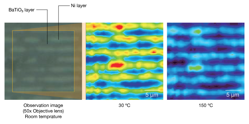
Color map by peak area at 520 cm–1
Surface Imaging – SSI
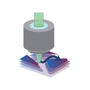
This method is useful for samples with uneven surfaces and the varying heights. This technique involves scanning the stage in three dimensions based on the sample height information obtained from the omnifocal image, without requiring any sample pretreatment. The spectrum is measured during scanning process.
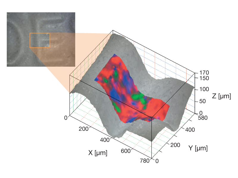
Engraved groove of pharmaceutical tablet
(red: etenzamide, green: acetaminophen, blue: caffeine)
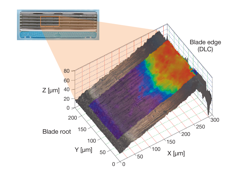
DLC coating distribution on razor edge
3D imaging
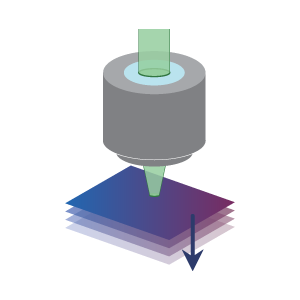
This method is useful for nondestructive and noncontact analysis of each layer in multilayer films or buried foreign matter. The confocal optical system enables Raman imaging of the sample’s depth to be obtained.
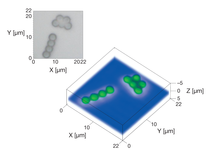
Polystyrene particles on Si wafer
(green: polystyrene particle, blue: Si)
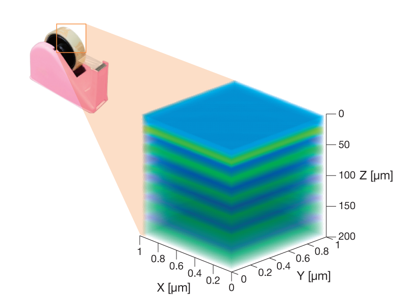
Scotch tape
(blue: substrate layer, green: adhesive layer)
