Introduction
<Question>
How can you easily measure micro foreign object by micro ATR?
<Answer>
By using of a “Clear-View” ATR objective, you can easily carry out the measurement under observation of sample image through the ATR prism. Especially in the measurement of micro foreign object in gel that the actual target position will move easily from the center of prism, the “Clear-View” ATR in the combination of IQ Mapping enables the measurement outside of center of prism area in contact with the sample.
In addition to the ordinary observation mode, the Model ATR-5000-SS(ZnS)/SD(Diamond)/SG(Ge*) ) “Clear-View” ATR objectives allow the users to observe the sample image through ATR prism in contact with the sample (See Figure 1).
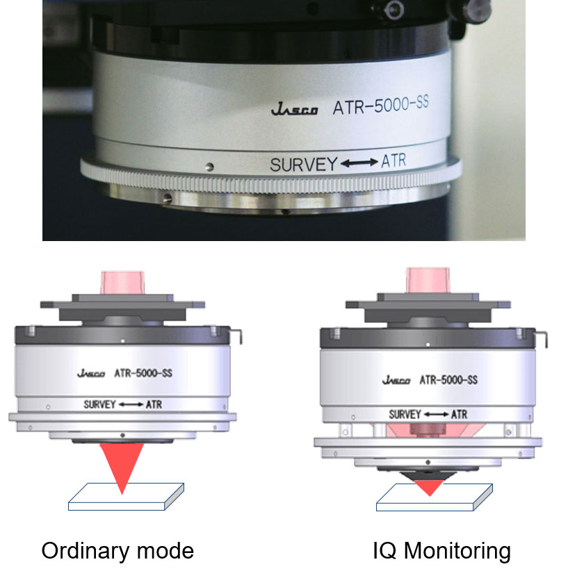
Figure 1. Clear-View ATR (ATR-5000-SS)
By attaching the “Clear-View” ATR objective to the IRT-5000/7000 Infrared Microscope, the IQ Monitoring (spectral measurement through the prism in contact with sample) and IQ Mapping (spectral measurement of outside of center of prism area by scanning of light axis.) can be performed. By utilizing of these new sampling modes, you can set the exact sampling point even in the measurement of micro foreign object in gel having high liquidity that the actual target position will move easily from the center of prism. Also the new modes can be applied to soft sample that the shape may change in contact with the prism or the ruggedness sample that is not easily in contact with the prism.
Also IQ Mapping offers contamination-free ATR mapping even in the measurement of sticky sample, because the mapping can be performed by scanning of light axis and not driving of stage. Therefore, the prism contacts the sample only once per one mapping.
*) ATR-5000-SG offers the ordinary observation mode only.
Experimental
Application of Clear-View ATR (ATR-5000-SS) – colored particle in nail polish –
The nail polish is a gel sample including several kinds of colored particles. Therefore, it can be the good sample to demonstrate the performance of Clear-View ATR. The ordinary observation image and the image in IQ Monitoring are shown in the Figure 2.
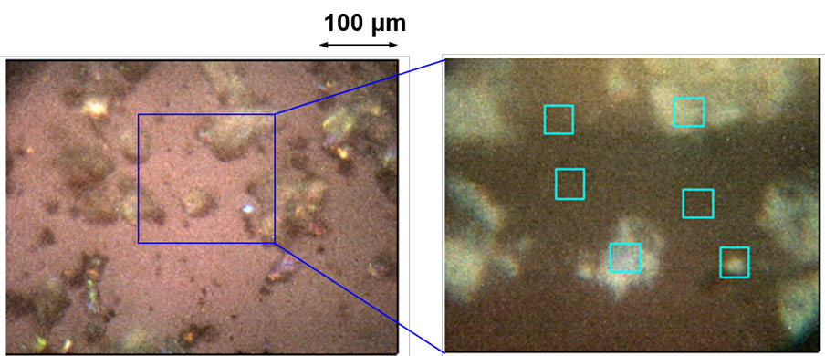
Figure 2. Ordinary observation image (left) and image in IQ Monitoring (right)
As you can see, the target sample, high light (particle) is positioned outside of center of prism in contact with sample. The multiple points measurement around the center of prism in contact with sample were carried out by IQ Mapping. The results indicate that base part is of dye and high light part is silica gel (See Figure 3.).
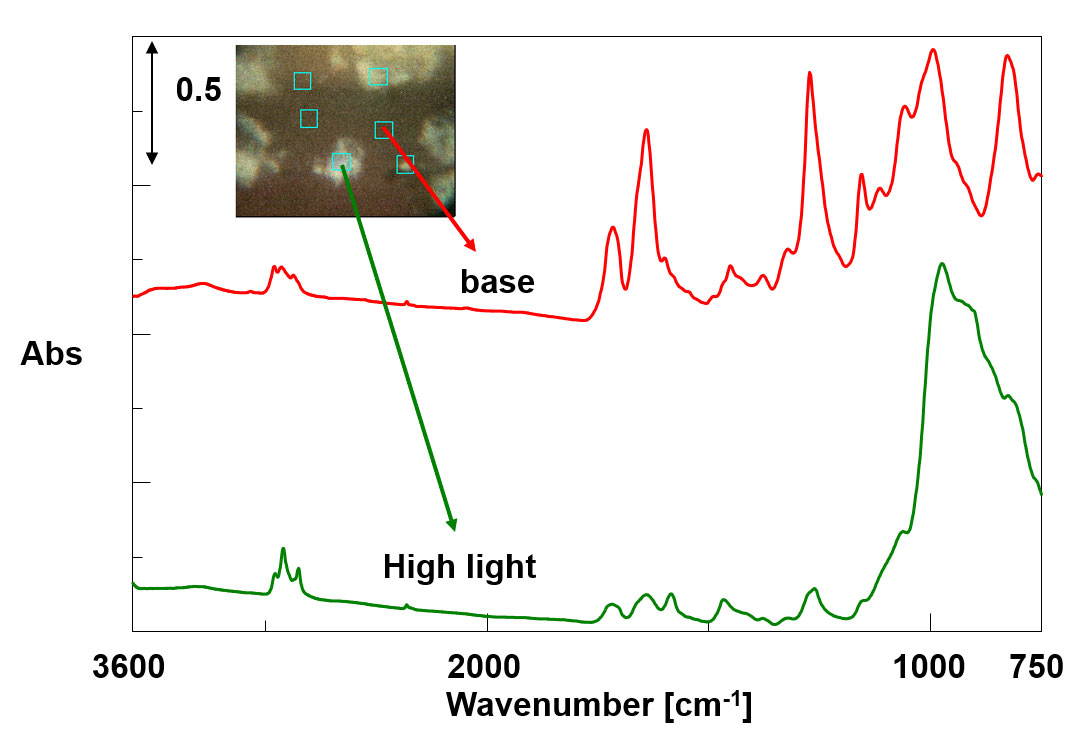
Figure 3. IR Spectra of nail polish

