Introduction
In infrared (IR) spectroscopy, the appropriate measurement technique is determined based on the sample shape. In recent years, the ATR method, which is applicable to a wide range of sample shapes, is commonly used. This method basically requires no sample dilution, thin-film formation, or other pretreatment, and measurements can be performed simply by pressing a sample against a crystal. It is applicable to solid, liquid, and powder samples.
However, before performing measurements using an ATR unit in the FTIR sample compartment (macro-ATR method), images are generally taken of microplastics or foreign samples using an optical (stereo) microscope in order to record their shapes and colors. The sample is then moved onto the ATR crystal, which takes a lot of time and effort. In addition, since the images and the spectrum are saved independently, they must be subsequently correlated, which is also time-consuming. To solve these problems, a macro-ATR unit equipped with a USB microscope has been developed. It takes before FTIR measurements while the sample is already on the ATR crystal.
Macro-ATR unit with USB microscope
- The USB microscope can be used with a macro-ATR unit such as the ATR PRO 4X VIEW or ATR PRO 4X.
- Images taken with the USB microscope can be viewed and saved in measurement and analysis programs.
- The obtained spectrum and images of the sample on the ATR crystal (only when the ATR PRO 4X VIEW is used), together with images of the sample taken with the USB microscope can be saved in a single file.
- This offers advantages when measuring small samples because the sample can be set on the ATR crystal while viewing the USB microscope image.
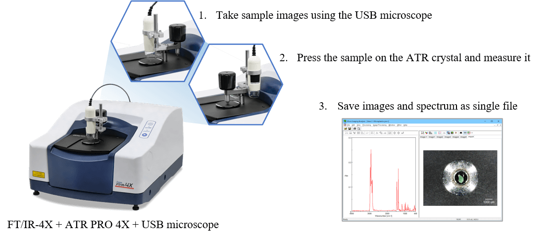
Fig. 1 Macro-ATR measurement system with USB microscope
<Collection location for microplastics>
Sample (1): Lake Biwa (Japan)
*Provided by Biwako Environmental Project 2021, sponsored by The Association of the Global Science Education
Sample (2): Shonan coast (Japan)
Keywords
Microplastics, Foreign substances, ATR method, Images of samples, USB microscope
Results
Figure 2 shows spectra acquired after taking sample images using the USB microscope mounted on the macro-ATR unit. The ATR PRO 4X VIEW, which is capable of acquiring images when the sample is pressed on the crystal, was used. Therefore, images of the sample pressed on the crystal before ATR measurements were also captured.
The images provided information on the shape, size, and color of the microplastics soon after they have been collected. Sample 1 is a very small sample with a long axis of about 200 μm, but observation with the USB microscope or the ATR PRO 4X VIEW allows for secure placement and measurement of samples on the crystal. A database search for the acquired spectra identified sample 1 as polypropylene (PP) and sample 2 as polyethylene (PE).
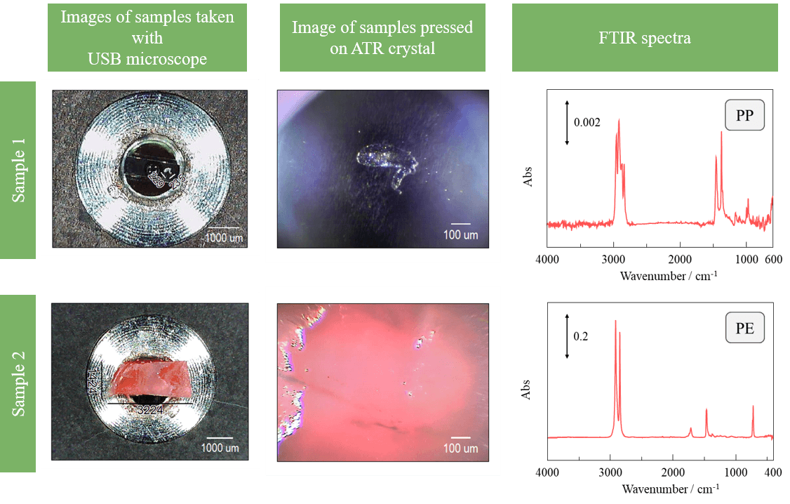
Fig. 2 Obtained images and spectra
Conclusion
The macro-ATR unit equipped with a USB microscope allows information about sample shape and color to easily be saved before measurement. Since the sample can be set on the crystal while viewing the image magnified by the USB microscope, and the acquired images and spectrum can be saved in a single file, an improvement in work efficiency is expected. The USB microscope can be conveniently used for ATR analysis of microplastics and foreign materials.


