Introduction
Infrared (IR) microscopy and Raman microscopy are commonly used to measure particle samples captured using a membrane filter. However, the membrane filter must be firmly secured to avoid wrinkling. Teflon (PTFE) membrane filters are often used for these analyses because of their wide variety of filter and pore diameters, low cost, and low number of spectral peaks that interfere with sample peaks in IR and Raman measurements. However, because a PTFE membrane filter is usually soft and it wrinkles easily during drying, it is difficult to firmly secure the filter for spectroscopic measurements without wrinkles being present. To solve such problems, a new and improved membrane filter holder for microscopic measurements (SH-02-FH Filter Holder), which allows a PTFE filter membrane to be easily held without wrinkles, has been developed for spectroscopic measurements. In this report, the use of the holder and measurement results for plastic particles on a PTFE membrane filter using IR and Raman microscopes are described.
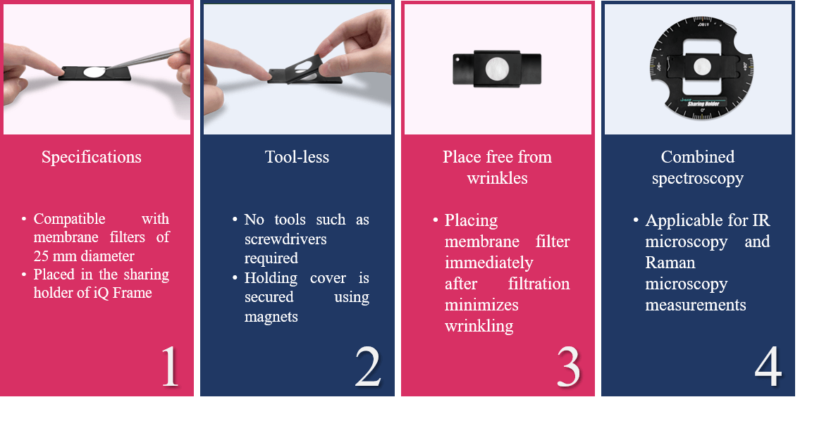
Fig. 1 Features of membrane filter holder for microscopic measurements
Experimental
<Procedure>
1.Plastic particles*1 were dispersed in water and collected on a PTFE membrane filter by suctioning filtration.
2.The filter was placed on the membrane filter holder for microscopic measurements immediately after filtration.
3.After drying, mapping/imaging measurements of the same area were performed independently, using IR and Raman microscopes.
The measurement area was approximately 1 mm x 1 mm, and iQ Frame*2 was used for aligning the measurement position between microscopes.
*1: Plastic particles were provided by TOSOH Analysis and Research Center Co., Ltd.
*2: Supplemental data: FT/IR application data 100-MT-0253, Raman application data 100-AN-0016
Keywords
Microplastics, Foreign substances, Small samples, Imaging measurement, iQ Frame, Filter holder
Results
Membrane filters could be maintained in a less wrinkled state by the newly developed holder, and it was found that imaging measurements over a wide area were possible. In this study, a 1 mm x 1 mm area was measured, but larger areas can also be measured.
Figure 2 shows imaging measurement results for the same area using IR and Raman microscopes. In this example, polyethylene terephthalate (PET), polypropylene (PP), polyethylene (PE), and polystyrene (PS) were identified. A chemical image (color-coded diagram) can be created based on the heights of peaks that are unique to each material and that are indicated by arrows in the spectrum. The results show that high spatial resolution Raman microscopy is capable of identifying the detailed particle distribution, and can detect smaller particles than IR microscopy. However, IR microscopy measured tens of times faster than Raman microscopy. By combining the advantages of both forms of microscopy, the appropriate analysis for the purpose can be achieved.
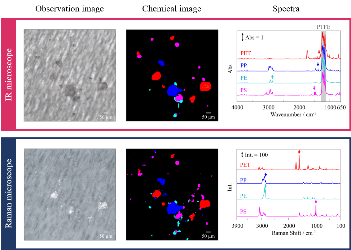
Fig. 2 Peak height plot based on peaks for each material
Conclusion
The newly developed filter holder allows the membrane filter to be easily secured and the sample particles to be measured. This new holder can be conveniently used for the analysis of microplastics and foreign substances captured on membrane filters.
Related Posts:
 Imaging measurement of plastic particles using IQ Frame
 Use of Diamond Compression Cell for Different Sample Types
 Optical Rotation Measurement for High-concentration…
 Stability Evaluation of Trastuzumab and Rituximab…
 Base Material and Dye Analysis - Combined Raman and…
 Highly efficient spectral measurement methods using…