Laser selection
Selection of the laser wavelength is important for obtaining meaningful Raman data. The excitation wavelength can be selected from the UV though the visible to the near IR regions, with a range of power options depending on the application. Multiple lasers with a variety of wavelengths can often be installed in a Raman microscope to allow data to be measured for different types of samples. A popular laser wavelength is 532 nm because it offers a relatively high Raman excitation energy and a low fluorescence intensity.
However, for a greater reduction in fluorescence, lasers with longer wavelengths such as 785 nm and 1064 nm are often used. Less frequently required UV lasers offer the additional benefit of “resonance Raman”, an excellent choice for carbonaceous materials.

Fig. 7 Laser selection for Raman spectroscopy
Fluorescence
Fluorescence can be a side effect in Raman measurements, and can originate from either the target molecules or the surrounding matrix. Care must be taken if the sample is contained in a vial or tube as this can also cause background interference. There are two physical methods for reducing fluorescence. The first is to select a laser wavelength at which fluorescence does not occur. This is typically a longer wavelength where there is insufficient energy for electron excitation. The second method is to choose the aperture size and shape to mask as much of the sample matrix as possible to eliminate matrix fluorescence – this is improved with dual spatial filtration (DSF) included with NRS-5000/7000 spectrophotometers. A third option is the use of a fluorescence rejection algorithm (patented), which is highly effective at removing the broader fluorescence spectrum, leaving the sharper Raman peaks with a clean baseline, as shown below. This feature can be used either automatically during measurement for fast imaging, or post-acquisition for additional data processing. This often works well for samples with strong fluorescence when using shorter-wavelength lasers (such as 405 or 457nm), which produce a more intense Raman signal.
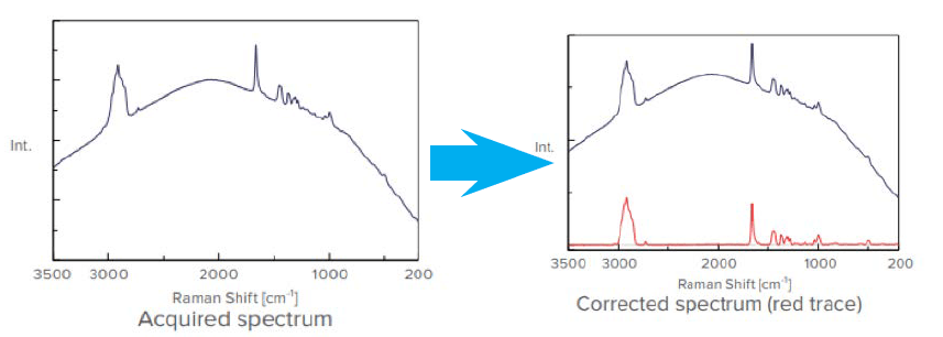
Fig. 8 Fluorescence correction can be performed during or post acquisition
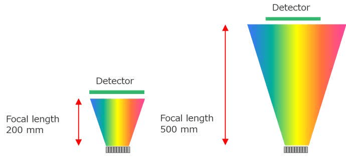
Fig. 9 Schematic showing dependence of dispersion on focal length
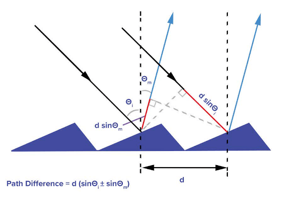
Fig. 10 Angular dispersion
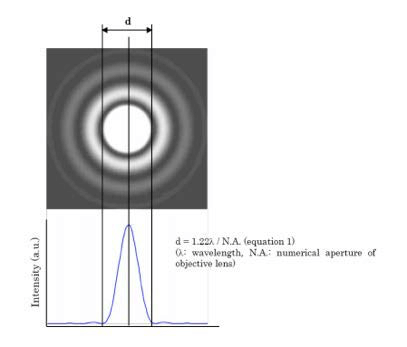
Fig. 11 Airy-disk resulting from diffraction with a circular aperture
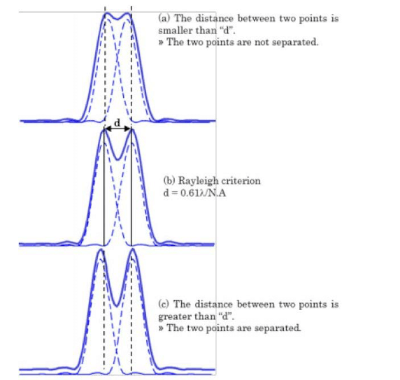 Fig. 12 Spatial resolution defined by the Rayleigh criterion
Confocal optics
The spot size in laser Raman microscopy is typically less than 1 μm. The optimized confocal design of the NRS- 5000/7000 includes an additional feature - dual spatial filtration (DSF), making it possible to achieve a maximum spatial resolution. DSF eliminates stray light that is not removed at the first aperture and significantly improves the resolution along the Z axis (Fig. 7).
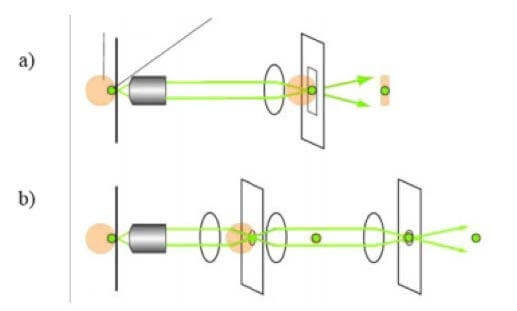
Fig. 13 a) Standard confocal optics with pinhole aperture; b) JASCO DSF(Dual Spatial Filtration) confocal optics
Related Posts:
 Avoiding Interference from Fluorescence Signal by…
 Analysis of pollen collected by Durham sampler…
 Introduction of New and Improved Sample Holder for…
 Use of Diamond Compression Cell for Different Sample Types
 Resonance Raman Scattering for Trace-Level Component…
 Structural Analysis of Chiral Samples with Multiple…