Advantages of Raman spectroscopy
Advantages of Raman Sampling
Raman spectroscopy offers a number of significant advantages over other spectroscopic or optical microscopy techniques. It can be used for depth profiling and microscopic area mapping of samples with a spatial resolution of around 1 μm. Optical fibers with remote probes can be used for sensitive in-situ analysis. With no combinations or overtones, there is less ‘clutter’ in Raman spectra. The technique is not sensitive to the presence of water or environmental vapor or gas. The measurement range is not limited by optical components, cells or accessories. An entire spectrum can be measured simultaneously in a wide range from 4,000 to 50 cm-1 (or even less) using a spectrometer with a CCD detector. Measurements can be performed on opaque or cloudy samples, and directly through glass or plastic containers, making sample preparation much simpler.
Advantages of Raman Microscopy
Raman microscopy is a non-contact, non-destructive technique that requires little to no sample preparation. Samples can be measured with a spatial resolution as small as 1 μm, and depth profiling can also be easily performed on transparent samples. Using confocal microscopy, measurement can be made in the Z direction for transparent samples, providing a three-dimensional chemical image of a sample. There are several important factors to consider when performing Raman measurements, which have a significant bearing on the quality of the data that can be obtained. Here, we will provide some insights into how to perform good Raman measurements, what may be expected from the instrument, and the key elements that affect performance.
Raman applications
Raman spectroscopy can be used in a wide variety of applications, as shown in Table 1. These include not only traditional quantitative / qualitative analysis but also microscopic applications involving scalable imaging techniques depending on the properties of the target samples.
Table 1 Raman applications
| Field | Applications |
| Pharmaceuticals | Light stability of ciprofloxacin tablets, xanthine derivative tablets, quinolone delivative antibacterial tablets, theophylline hydrates, anhydrates, crystalline polymorphs of indomethacin, crystalline polymorphs of carbamazepine (CBZ), crystalline polymorphs of ampicillin, crystal structure and thermal stability of acetylsalicylic acid (aspirin), active ingredients in drug substances and their preparation (Jpn Pharmacopeia), qualitative/quantitative evaluation of additives (Jpn Pharmacopeia), bronchodilator (TBR, turobuterol) tape |
| Foods | Component distribution on white chocolate surface, butter/margarine emulsion imaging , components of egg yolk, thermal change of trehalose dihydrate, fatty acid in food oil, aaccharides solutions (saccharose, glucose, xylitol, galactose, lactose), multilayer films for food packaging, ethanol in glass bottles, caffeine, crystallinity of PET bottles |
| Carbon materials | Carbon nanotubes, diamond-like carbon, fullerenes |
| Semiconductors | Power semiconductor (SiC) devices, crystallinity of polysilicon |
| Electronic devices | Foreign matter in liquid crystal substrates, foreign matter in color filters, diamond-like carbon on hard disk surfaces, solar cells (crystalline silicon, amorphous silicon) |
| Polymer compounds | 3D imaging of cellophane tape, polypropylene-polyethylene multilayer films,foreign matter on polyethylene films, polymer additives, dispersion in blended polymers, crystallization of molten polymers, curing of UV curable resin, dispersion of lubricant on films, orientation of natural rubber, synthetic rubber |
| Biological materials | Visualization of sea-island structure in blended polymers, structural changes in proteins (hemoglobin, lysozyme, cytochrome c), enzymes (ribonuclease A), dental adhesive, collagen, chemical imaging of coral, structure and orientation evaluation of spider silk |
| Cosmetics | Ingredients of lipstick marks, eye shadow |
| Gas | Natural gas hydrates |
| Others | Imaging of bath powder (mixed powder samples), carbon nanotubes, crystallinity of core of pencils, identification of fingerprints with vermilion ink, iron rust, colored fibers, Nylon 6 fibers, wood (lignin), quartz, calcite, nondestructive analysis of archaeological material (mainly pigments) |
Raman imaging
Raman imaging is a powerful technique that provides 3-D spatial information and chemical identification (Fig. 6). Samples with dimensions of micrometers to millimeters can be analyzed in just a few minutes. JASCO has developed a technology called QRI that increases the data acquisition speed by up to 50 times compared with conventional mapping, and also offers a dramatic improvement in sensitivity.
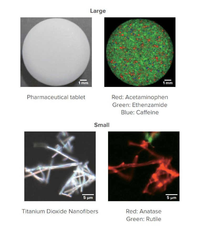
Fig. 6 Raman imaging for spatial distribution and chemical identification
Related Posts:
 Two-dimensional Correlation Spectroscopy for…
 Raman Measurement in Containers -A Simple…
 In-situ Raman Monitoring for Flow Synthesis of Peptide Drugs
 Can the JASCO Palmtop Raman spectrometer perform a…
 Imaging measurement of plastic particles using IQ Frame
 Analysis of Diquat – Methods for Testing Water…