Introduction
When measuring transmittance or reflectance of a sample that diffuses or refracts light, such as frosted glass or lens, an integrating sphere is generally attached to the spectrophotometer. Since the incident light usually travels horizontally in the sample compartment, the sample holder is designed so that the sample can be mounted vertically. However, samples with nonparallel surfaces or irregular shapes, such as teeth or contact lenses, are difficult to mount in this way. For this reason, JASCO has developed an integrating sphere that allows horizontal mounting of samples (Figs. 1 and 2) during measurements (Japan Patent No. 5925725).
In this report, we describe diffuse reflection measurements of a human tooth. The colors at different positions on the tooth were determined using color evaluation software. The results show that this integrating sphere is useful for samples with nonparallel surfaces.
Experimental
Sample
Human tooth
System
V-750 UV-visible spectrophotometer
PIV-756 Horizontal sampling integrating sphere
Lens unit for reflectance measurements
VWCD-960 Color evaluation – color diagnosis
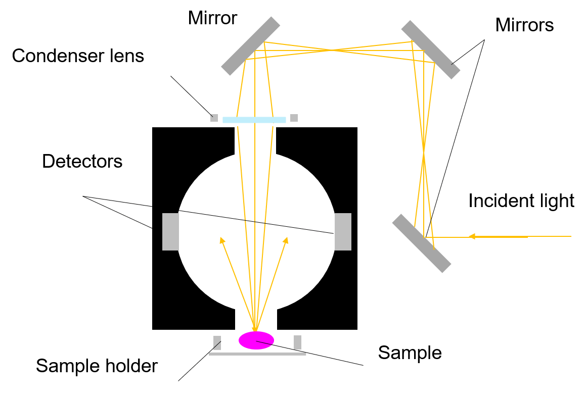 Fig. 1 Schematic diagram of PIV-756 (side view)
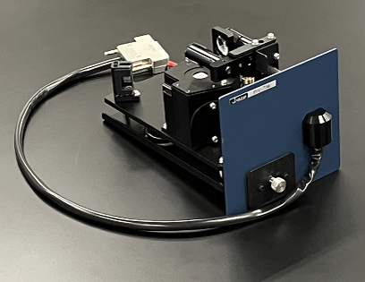 Fig. 2 PIV-756
Conditions
Measurement range: 380 to 780 nm
Data interval: 1.0 nm
Bandwidth: 5.0 nm
Response: 0.24 sec
Scanning speed: 400 nm/min
Keywords
Horizontal sampling integrating sphere, irregular shape, tooth, diffuse reflection, color evaluation
Results
Diffuse reflectance spectra of enamel and dentin in a tooth were obtained using the horizontal sampling integrating sphere. The spectra were measured at two different points for each material. A condenser lens and a ø1-mm mask, placed in front of the opening of the integrating sphere, were used to adjust the diameter of the incident light spot on the sample. The lens is movable by hand, which allows the light spot position to be adjusted without moving the tooth.
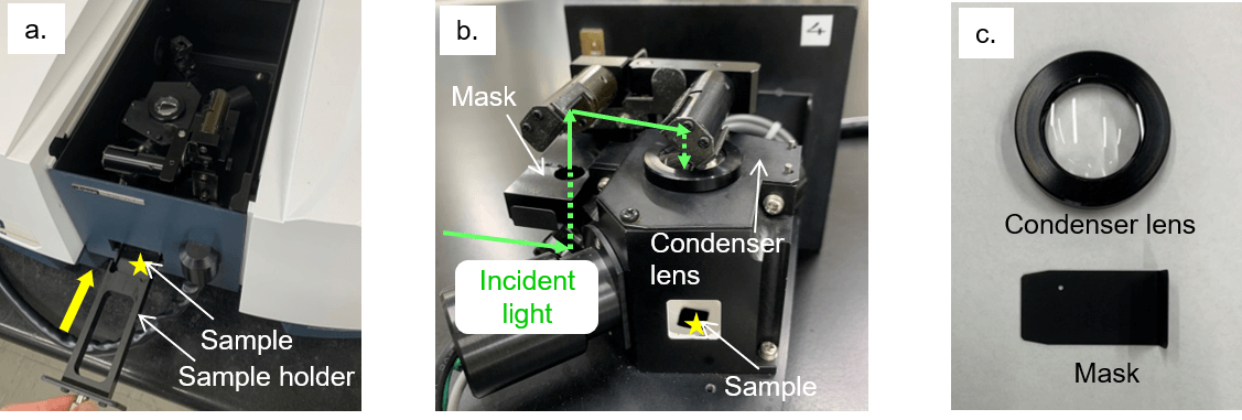
Fig. 3 Integrating sphere for horizontal samples: a) front view, b) back view, c) lens and mask
As shown in Fig. 4a, the spectra obtained for the same material were almost identical; however, different spectral shapes were obtained for the enamel and the dentin. Using the color evaluation software, the L*, a*, and b* values were calculated from each spectrum. The L* and a* values were almost the same for enamel and dentin, while the b* values were different, as seen in Fig. 4b. This difference indicates that the dentin is yellower than the enamel, which is consistent with the visual appearance of these materials.
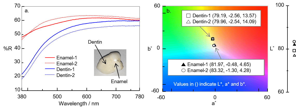
Fig. 4 Results: a) diffuse reflectance spectra, b) L*a*b* color chart
Conclusion
Diffuse reflectance spectra of a tooth, which has nonparallel surfaces, were successfully obtained using the horizontal sampling integrating sphere. The movable condenser lens allowed the incident light spot to be placed at the desired position without moving the sample. The color difference between the enamel and the dentin was statistically examined using color evaluation software. This application note shows that this integrating sphere is suitable for diffuse reflection measurements of samples that are difficult to mount vertically.


