Introduction
With the advent of multi-channel detectors, there have been significant advancements in imaging using infrared microscopy. In particular, microscopic imaging using attenuated total reflection (micro-ATR imaging) has attracted a great deal of attention as a surface analysis method because of the lack of need for sample preparation. In micro-ATR imaging, measurements are performed by placing a specimen and a prism in close proximity to each other, which limits the measurement area. However, wide-area measurements are possible by using a specially shaped prism. We have previously developed a Fourier-transform infrared spectrophotometer using micro-ATR imaging, in which the optical axis of the infrared light can be scanned. This allows a seamless transition between sample observations and measurements.
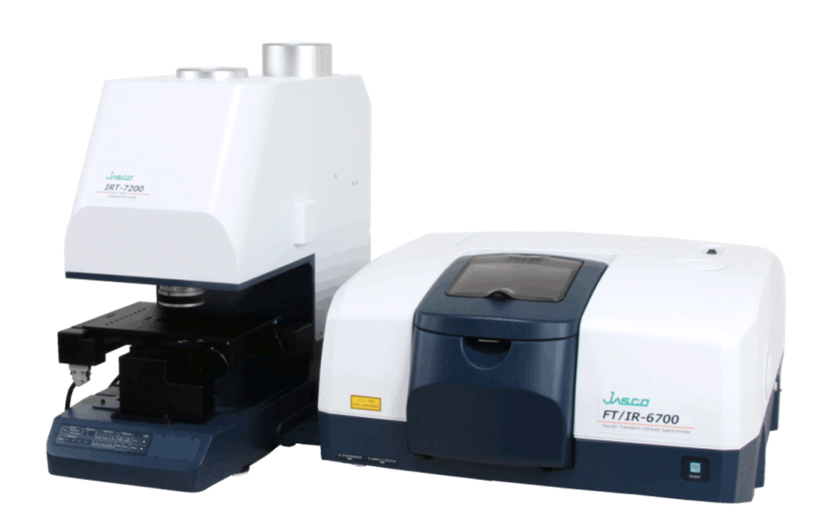
Micro FTIR system FT/IR-6700 + IRT-7200
IQ mapping using scanned infrared light
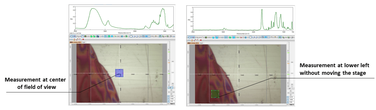
In general, when performing ATR imaging, the contact between the prism and the sample is broken when changing the measurement point, and after moving the stage, the contact is reestablished. Although this method allows wide-area measurements, there is a possibility of sample contamination. Therefore, we have developed a method called IQ mapping, in which the infrared light beam is scanned without moving the stage. This can be used even for ATR measurements, with no risk of contamination. Even for samples whose shape is likely to change due to contact with the prism, accurate imaging is possible.
Features of ATR-5000-WG for wide-area ATR imaging
By improving the optical system, we developed the ATR-5000-WG objective mirror for wide-area imaging. The table below compares the features of the ATR-5000-WG (WG), the ATR-5000-MG (MG) and the ATR-5000-G45 (G45).
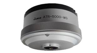
ATR-5000-WG appearance
| WG | MG | G45 | |
|---|---|---|---|
| Magnification | 4x | 16x | 90x |
| Prism material | Ge | Ge | Ge |
| Average incidence angle | 35º | 42º | 40º |
| Sample refreactive index | < 2.2 | < 2.4 | < 2.6 |
| IQ mapping | 1600 x 1600 μm | 400 x 400 μm | 70 x 70 μm |
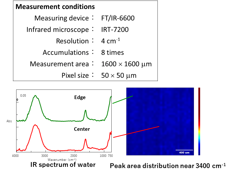
Basic data: water imaging
We performed imaging measurements of the sample (water) that was in intimate contact with the entire prism.
Since there was no change in the shape and intensity of the spectrum in all the measured regions, it can be said that ATR-5000-WG can measure a uniform spectrum in the entire 1600 × 1600 μm region.
Experimental
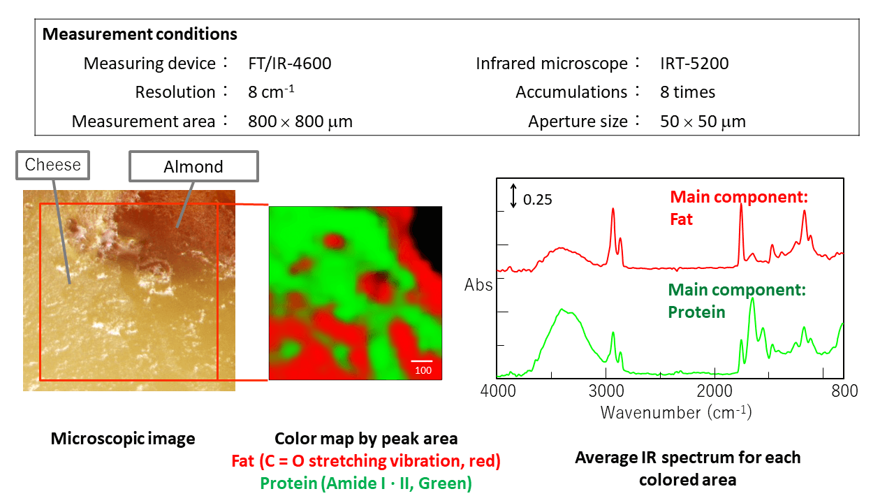
Measurement 1: Dairy product containing nuts
A cross section of cheese with almonds was measured by ATR imaging. Even for samples in which soft cheese and hard almonds are mixed, the sample shape and component distribution can be accurately measured by smart mapping with a single contact. The distribution of fat and protein can be evaluated using the ATR-5000-WG.
Measurement 2:Laminated functional material
A cross section of the paper beverage container was measured using the ATR-5000-WG, ATR-5000-MG and ATR-5000-G45. Using the ATR-5000-WG, the entire sample was measured, and the distribution of three components in six layers was evaluated. Using the ATR-5000-MG, the center of the sample was measured, and the polyethylene layer between cellulose layers could be detected as a result of the high spatial resolution. Using the ATR-5000-G45, a layer of trace calcium carbonate could be detected at even higher spatial resolution.

Conclusion
Conclusions
It was shown that the ATR-5000-WG was capable of high-quality imaging in a 1600 ´ 1600 mm region. Cheese containing almonds was measured to visualize the distribution of fat and protein.
A cross section of a laminated material was measured by ATR imaging using three types of ATR objectives, and the components in each layer could be evaluated.
The combination of IQ Mapping and the ATR-5000-WG can be used to evaluate soft materials such as polymer blends, pharmaceutical capsules, rubber, food and laminated samples.
References
Authors
Higuchi, Yuji ; Kanno, Miyuki ; Tamura, Kohei ; Watanabe, Keisuke JASCO Corporation
This application was introduced at ICAVS 10 in New Zealand.
Related Posts:
 Development and Applications of Full-Vacuum FT-IR…
 Orthogonal Assessment of Polymer Materials Including…
 Microplastics Identification using Macro-ATR Unit…
 Analysis of pollen collected by Durham sampler…
 Base Material and Dye Analysis - Combined Raman and…
 GPC/SEC measurement of Polystyrene Oligomers using…