Introduction
There are various devices that can create cross-sections of multi-layer films for analysis of the molecular structure using infrared spectroscopy. To date, cross sections of multilayer laminates have been created by embedding the sample in epoxy resin and then using a microtome to create the cross-section slices. However, this technique has various drawbacks, including the time required for the resin to harden (several hours for epoxy resin) as well as resin contaminating the sample. The SliceMaster can provide thin ‘slices’ or cross sections of various samples, including multi-layer laminates, without embedding them in resin. This article shows the measurement example of cross-section (food package sample) which the vertical SliceMaster (HS-1) (Figure 1) created.
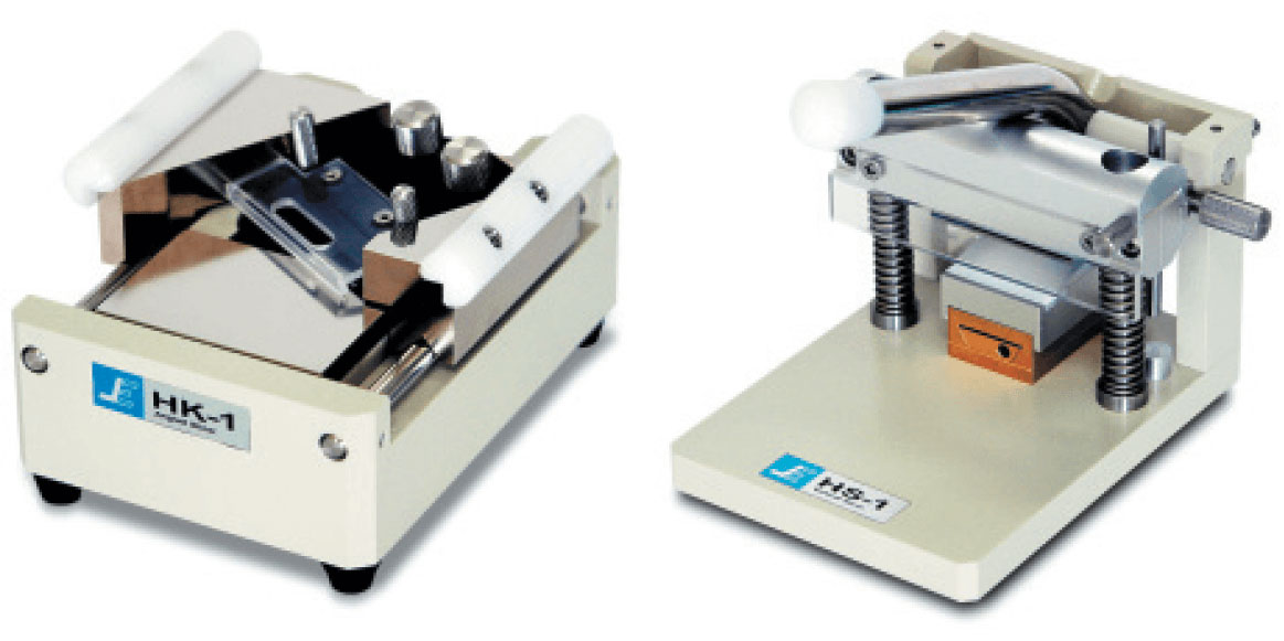
Figure 1. SliceMaster (left: HK-1 angled slicer, right: HS-1 vertical slicer)
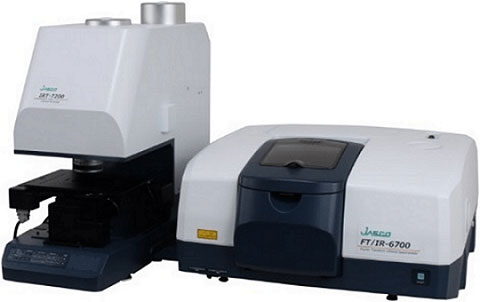
FTIR microscope system (FT/IR-6700 + IRT-7200)
Results
The measurement area was 600 × 600 μm with a spatial resolution of 12.5 μm, collecting 16 scans at 8 cm-1 resolution for all spectrum. Based on the spectra collected for each layer, we determined that the laminate was comprised of polyethylene (PE), polyvinyl alcohol (PVA) and poly ethylene terephthalate (PET) (Figure 2(1)). Figure 2(2) is the visual image of the sample captured with the integrated CCD video camera in the IR microscopy. Figure 2A is the infrared image based on the peak intensity at 3500 cm-1 attributed to the -OH functional group for the PVA material; Figures 2B and 2C are based on the peak intensity for the C-H stretching absorption at 2920 cm-1 and the C-H bend at 1440 cm-1, respectively, present in all the layers. Figure 2D represents the image based on the C=O peak intensity at 1730 cm-1, present in the PET layer, and Figure 2E is the image for the peak intensity of the 1250 cm-1 absorption. The infrared images obtained for the sample revealed the clear differences among the four-layer structure.
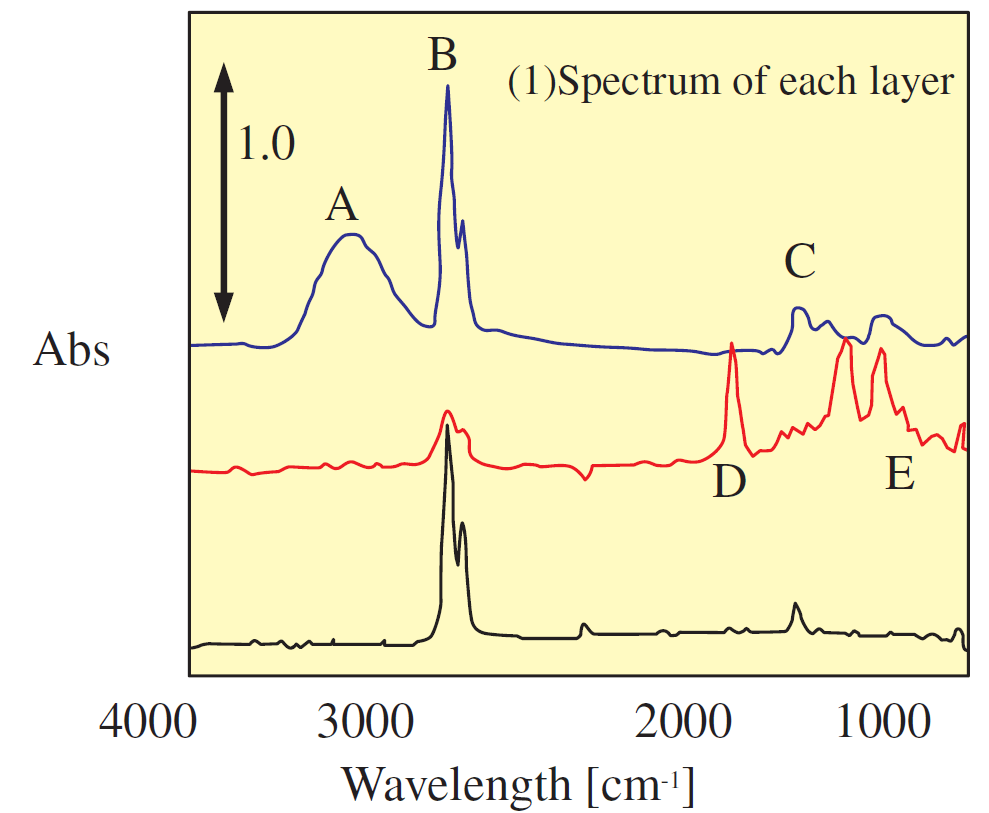
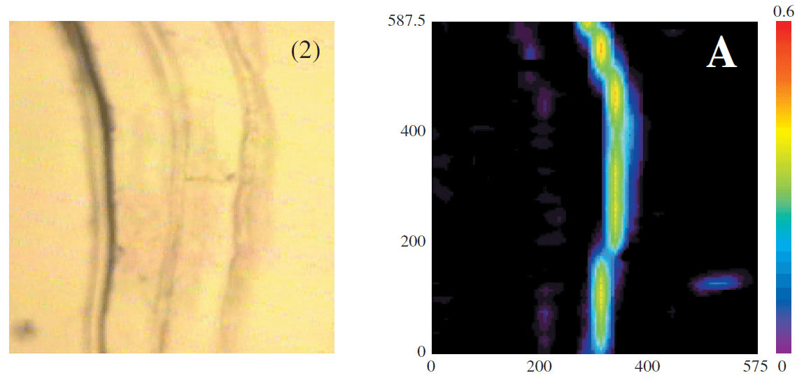
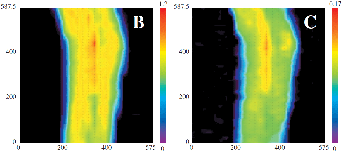
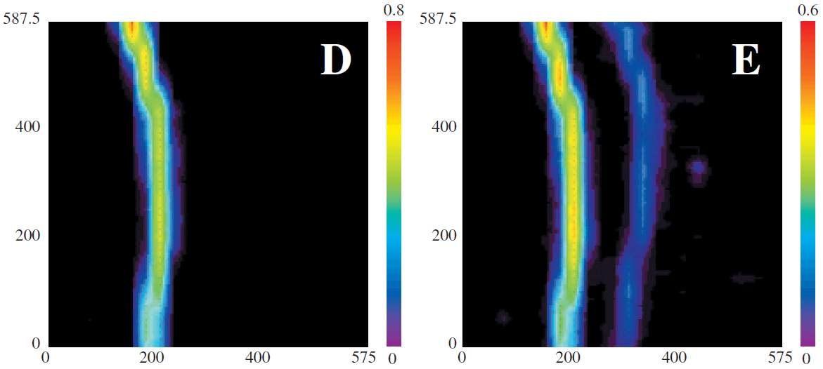
Figure 2. Multi-layer measurements
Based on these results, information on the adhesive layers between each film layer could not be obtained, but the use of the HK-1 angled SliceMaster and the 32× cassegrain objective, could provide the ability to obtain greater spatial resolution for analysis of the sample.
Related Posts:
 Identification of Degraded Plastics Cross-checking…
 Evaluation of Higher-Order Structure and…
 Imaging measurement of plastic particles using IQ Frame
 Introduction of New and Improved Sample Holder for…
 Use of Diamond Compression Cell for Different Sample Types
 Optical Rotation Measurement for High-concentration…