Introduction
Currently, the infrared microscope is used extensively as one of the identification approaches for micro foreign materials. Since in microscopic infrared spectroscopy there is a huge database, it work very well on the identification of foreign materials, while the infrared microscope has several problems for the measurement such that the spatial resolution is limited to only a few micrometer and the sample preparation is necessary if the foreign material is buried in the sample. Therefore, the Laser Raman spectrophotometer is now often used for measurement of foreign materials in combination with infrared microscope.
Raman spectroscopy is a method to analyze molecular structure by molecular vibration as well as infrared spectroscopy, but there are following advantages in Raman spectrophotometer.
- The spatial resolution is as small as 1 µm by using visible laser.
- The Raman spectrophotometer allows quick and easy measurement of the sample with non-destructive manner without sample pretreatment.
- For inorganic samples, it is easy to identify because of the easy measurement in low wavenumber range.
The potential of measuring foreign materials by using Raman spectrophotometer is expanding, and JASCO can provide laser Raman spectrophotometer with compact design and ease of use to be used together with FTIR. This article shows the identification example of foreign materials buried in the polymer film by using Raman spectrometer.
NRS-4500 Raman spectrometer
Experimental
The foreign material buried in the multi layer substrate (Glass/Adhesion layer/Transparent film) shown in Figure 1 was measured. It is difficult to measure such foreign material by using infrared microscope, because it is difficult to cut the foreign material in section due to the presence of glass and the adhesion layer may be picked up together. On the other hand, Raman spectrophotometer with the confocal optical system can obtain the spectrum of laser focused point selectively. As a result, it is possible to measure the inside of the sample in non-contact and non-destructive manner without lousy sample pretreatment. In this report, the position where the target foreign material is located was measured in the depth direction (Z axis direction) and also each layer’s information was obtained.
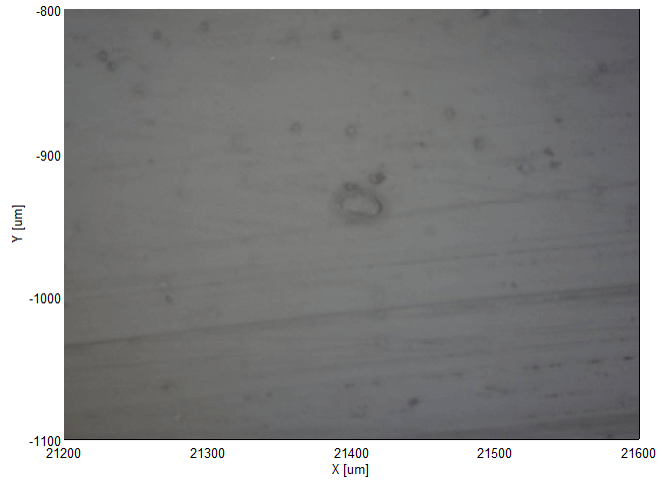
Figure 1. Observation image
Measurement parameters
Ex wavelength: 532 nm
Grating: 900 gr/mm
Exposure time: 5 sec. (Accumulation: 2 times)
Results
The spectra obtained from each layer are shown in Figure 2. In the obtained spectrum of the foreign material, the C-H peak at around 3000 cm-1 was not shown, and so it is quite different from the spectrum of transparent film and adhesion layer.
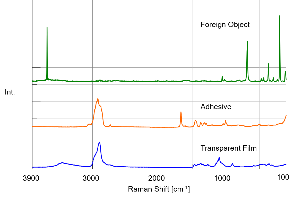
Figure 2. Spectra for each layer
In order to analyze the result in further details, the spectrum of foreign material was tried to be identified by using database as shown in Figure 3, and the foreign material was found to be talc (hydrated magnesium silicate). In addition, it is known that the transparent film is made of cellulose and the adhesion layer is made of terpene resin.
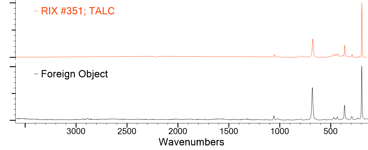
Figure 3. Search result of the database
Conclusion
Raman spectrometer can measure the buried sample in non-contact and non-destructive manner without lousy sample pretreatment. By using Infrared microscope together, the analysis of more complex foreign materials can be done.


