Introduction
< Key Points >
Raman spectroscopy provides a powerful approach for detecting trace amounts of components such as pigments.
Raman spectroscopy is an advanced analytical technique that enables the acquisition of molecular vibrational information from a sample in a non-destructive and non-contact manner by irradiating the sample with a laser and detecting the scattered light. In general, the detection of target components by conventional Raman spectroscopy requires their presence at concentrations of percent order. However, there are many analytical applications where the identification of components present at much lower concentrations is required.
For example, in the analysis of pigments contained in colored plastics, the pigment content is typically as low as approximately 1%, while the remaining ~99% consists of polymer resin. As a result, the Raman scattering signal originating from the resin is overwhelmingly strong, causing the spectral information of the pigment to be masked and making its detection difficult under conventional measurement conditions.
In this application note, a case study is presented in which trace pigment components contained in colored resins, normally difficult to detect using standard Raman spectroscopy, are selectively detected by exploiting the phenomenon of resonance Raman scattering.
What is Resonance Raman?
Resonance Raman scattering is a phenomenon that occurs when the energy of the excitation wavelength is close to that of an electronic transition of the target component. Compared with conventional Raman scattering, the signal intensity can be significantly enhanced, in some cases by more than four orders of magnitude. As a result, selective detection of specific trace components may be achieved by appropriately choosing the laser excitation wavelength used for measurement.
It should be noted, however, that the sensitivity enhancement associated with resonance effects occurs only for specific vibrational modes. Consequently, resonance Raman spectra may differ in spectral profile from those obtained under non-resonant conditions.
Examples of applications exploiting this phenomenon include the detection of trace amounts of carotenoids and/or polyenes1) present in biological samples such as egg yolk and green-yellow vegetables, as well as the selective analysis of carbon nanotubes with different structures.
Fig. 1 NRS-4500 Raman spectrometer
Experimental
A yellow-colored resin was analyzed using the NRS-4500 laser Raman spectrometer, featuring support for up to three laser wavelengths, with excitation such as 457 nm, 532 nm, and 785 nm (Fig. 1).
Sample
Yellow-colored polyamide pellet
Non-colored polyamide pellet
Yellow pigment
System
Instrument: NRS-4500 Raman spectrometer (Fig. 1)
Parameters
Excitation wavelength: 457, 532, 785 nm
Objective lens: 100x
Exposure time: 2-15 sec
Accumulation: 2 times
Keywords
Raman spectroscopy, resonance Raman scattering, trace amount component, pigment, dye, conjugate structure
Results
As a reference, uncolored polyamide pellets and a pure pigment sample were first measured using a 457 nm laser (Fig. 2). In both cases, well-defined spectra were obtained, and the spectral profiles were clearly distinct. The polyamide spectrum exhibited prominent bands attributable to hydrocarbon vibrations in the 3000-2800 cm-1 region and at 1446 cm-1, whereas the pigment spectrum showed a strong peak at 1459 cm-1.
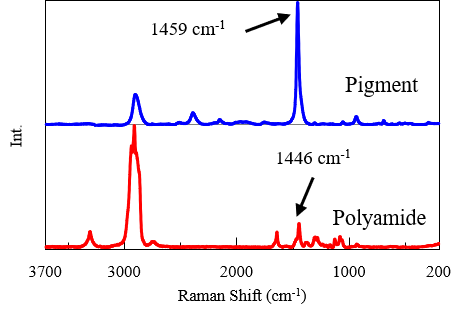
Fig. 2 Spectra of pigment and non-colored polyamide using 457 nm laser
Subsequently, yellow-colored polyamide pellets were measured at three different excitation wavelengths (457 nm, 532 nm, and 785 nm). In principle, Raman scattering is independent of excitation wavelength, and spectra with identical profiles would be expected. However, in this case, three spectra with markedly different profiles were obtained depending on the excitation wavelength (Fig. 3). When using the 785 nm laser, bands were observed in the 3000-2800 cm-1 region and at 1446 cm-1, and the obtained spectrum closely resembled that of polyamide shown in Fig. 2. In contrast, excitation at 457 nm produced a spectrum dominated by a strong peak at 1459 cm-1, which closely matched the pigment spectrum. At 532 nm, the spectral profile appeared to be a summation of those obtained at 785 nm and 457 nm.
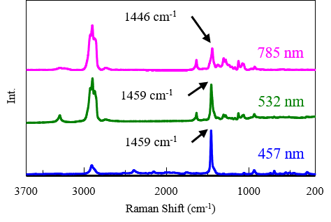
Fig. 3 Comparison of spectra of yellow-colored polyamide measured by three different wavelength
Conclusion
By exploiting the phenomenon of resonance Raman scattering, information on pigments present in trace amounts within the resin, normally difficult to detect, could be obtained. Furthermore, by carefully combining measurements with multiple laser wavelengths, the spectral features of the resin itself could also be selectively acquired.
Resonance Raman scattering generally occurs more readily in compounds with conjugated structures, making it a powerful tool for the detection of trace components such as pigments, as well as carotenoids in foods and biological samples.
References
1.M. Tsuboi, K. Tamura, R. Kitanaka, H. Oka, K, Akao, Y. Ozaki: Appl. Spectrosc., 78, 186 (2024). DOI: 10.1177/00037028231219026


