Introduction
< Key Points >
Melting experiments at temperatures above 95 ºC using an air-cooled Peltier device cell holder without a circulating thermostatic bath
Automation of the entire process from annealing to melting-temperature measurement
< Introduction >
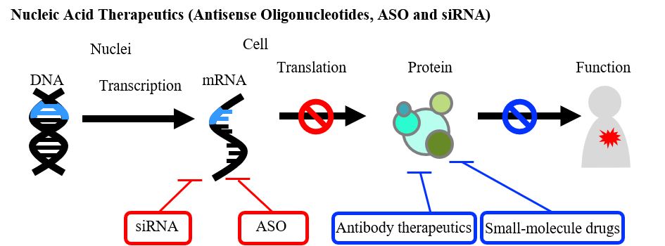
Nucleic acid therapeutics, composed of oligonucleotides typically consisting of about a dozen to several dozen bases, have attracted significant attention as a new drug modality. In recent years, many nucleic acid–based drugs have been launched1). Antisense oligonucleotides and small interfering RNAs (siRNAs) can regulate gene expression in a sequence-specific manner, allowing them to target “undruggable” proteins that cannot be approached by small-molecule or antibody therapeutics2, 3). Antisense oligonucleotides form double strands with the sense strand of the target mRNA, leading to mRNA degradation or exon skipping to control protein expression2, 3). In contrast, siRNAs are incorporated into the RNA-induced silencing complex (RISC) in their double-stranded form and induce cleavage of pre-mature mRNA to suppress target protein expression2, 3). Because duplex formation is essential for their biological activity, the melting temperature (Tm), which reflects duplex stability, is an important psychochemical parameter. In melting experiments to determine the Tm value, the sample is typically heated to around 95 ºC. Therefore, a water-cooled Peltier device cell holder with a circulating cooling bath is generally used to protect the Peltier element. JASCO offers the ETCS-761/762 single-cell Peltier device holder and the PAC-743 Peltier device cell changer, both water-cooled, with the latter capable of automatically measuring up to eight samples.
With the expansion of the nucleic acid therapeutics market, there is growing demand for compact systems that allow high-temperature measurements without the need for additional equipment such as a circulating thermostatic bath.
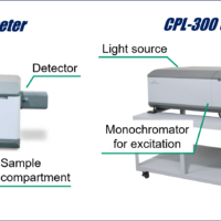
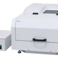
To address this demand, JASCO has developed the EHCS-932 single-cell Peltier device cell holder, which allows heating above 95 ºC without an external cooling unit, as well as an automated measurement system that performs both annealing and Tm evaluation (Figure 1).
This application note introduces this new system and describes its ability to precisely determine the Tm and other thermodynamic parameters for duplex formation of oligonucleotide
Experimental
Oligonucleotide (ODN)
Two complementary oligonucleotides were dissolved in 20-mM sodium phosphate buffer (pH 7.8) containing 100 mM KCl to prepare a sample solution with a total oligonucleotide concentration of 4 µM.
ODN1: 5’-CGC CCC CGC CCC CGC CCC GCC CCG-3’
ODN2: 5’-CGG GGC GGG GCG GGG GCG GGG GCG-3’
System
<Circular dichroism (CD) experiment>
Instrument: J-1500 CD spectrometer
Accessory: PTC-510 Peltier-thermostatted single cell holder
CTU-100 Circulating thermostat unit
<UV melting (Tm) analysis system>
Instrument: V-730 UV-Vis spectrophotometer
Accessory: EHCS-932 Air-cooled Peltier-thermostatted cell holder
Software: VWTP-959 Temperature control program
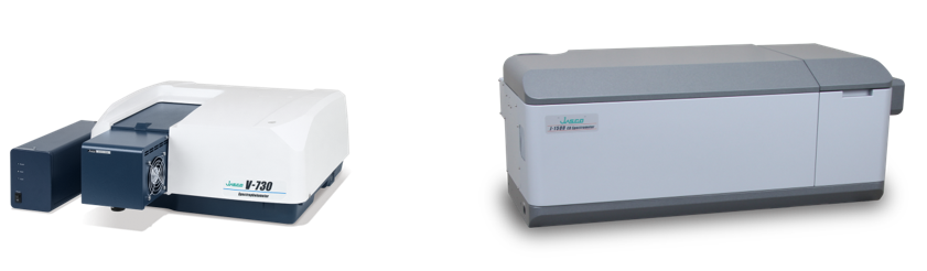
Fig. 1 UV/Vis Tm analysis system / CD spectrometer
Parameters
<Circular dichroism (CD) experiment>
Measurement range: 2200 nm – 320 nm
Scan speed: 50 nm/min
Bandwidth: 1.0 nm
Response: 4 sec
Data interval: 0.1 nm
Accumulations: 4
Path length: 5 mm*
<UV melting experiment>
Monitor wavelength: 260 nm
Temperature range: 25 ºC – 95 ºC
Response: 0.96 sec
Bandwidth: 1.0 nm
Path length: 5 mm*
Temperature monitor: Cell
Sample volume: 400 mL
*The same rectangular quartz cuvette was used for both the CD spectral and UV melting measurements to ensure a seamless transition between the two experiments.
Annealing
The sample solution was heated to 95 ºC and maintained at that temperature for 15 min, followed by cooling to 25 ºC at a rate of 2 ºC /min.
Keywords
Nucleic acid therapeutics, Oligonucleotide, Antisense, ASO, siRNA, Aptamer, Duplex, Tm, Melting temperature
Results
UV Melting Curve and CD Spectrum for Non-Annealed Oligonucleotides
To evaluate the necessity of the annealing process, CD spectra and melting curves were obtained before and after annealing using the procedure shown in Figure 2. CD spectroscopy is widely recognized as a powerful technique for characterizing the higher-order structures of nucleic acids4). In this study, the same rectangular quartz cuvette was used for both the CD and Tm measurements, allowing seamless switching between the two experiments.
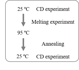
Fig. 2 Measurement procedure
The UV melting curve obtained without annealing exhibited a complex, non-sigmoidal profile, suggesting the presence of overlapping melting transitions originating from higher-order structures other than the intended duplex form (Figure 3a).
The CD spectrum of the non-annealed sample showed a shoulder at around 280 to 300 nm, indicating the coexistence of various structures such as self-complementary strands and quadruplexes in equilibrium, in addition to the duplex form (Figure 3b) 5–7).
In contrast, the CD spectrum obtained after annealing showed a reduced shoulder around 280 to 300 nm, suggesting that the annealing process effectively decreased the population of non-duplex structures.
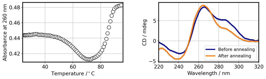
Fig. 3 UV absorption and CD results for oligonucleotides.
a) UV melting curve for non-annealed sample, b) CD spectra before and after annealing
Melting Experiment Using Automatic Annealing
The automatic annealing feature integrated into the [Temperature Control] program for the JASCO spectrophotometer was used to perform both the annealing and melting experiments in a fully programmed sequence (Figure 4). The melting curves obtained from three independent measurements all exhibited a clear sigmoidal profile (Figure 5).
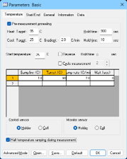
Fig. 4 Parameter settings
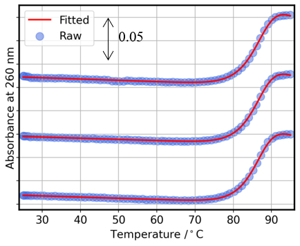
Fig. 5 UV melting curves
Subsequent fitting of these curves using the [Melting Analysis] program (Figure 6) with a non-self-complementary strand model (equations 1 and 2) yielded highly consistent Tm and ΔH values, demonstrating excellent reproducibility from measurement to analysis.
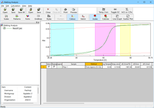
Figure 6. [Melting Analysis] program window
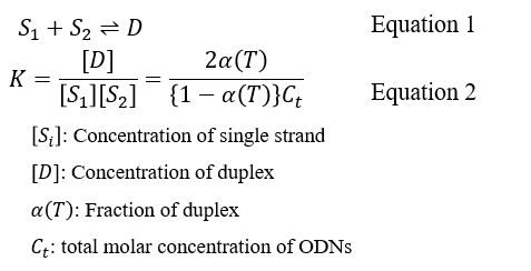
Table 1. Calculated thermodynamic parameters
| Ave. | S.D. | |
| Tm/°C | 86.4 | 0.16 |
| ΔH/ kJmol-1 | -615 | 11 |
Conclusion
By combining the automatic annealing function with a compact system that requires no circulating thermostatic bath, the melting temperature for oligonucleotides can be measured easily and with high precision. Furthermore, when coupled with CD spectroscopy—a powerful technique for evaluating higher-order nucleic acid structures—this system enables detailed characterization of duplex formation in oligonucleotides under development.
References
1.Editorial: Nat. Rev. Mater., 6, 99 (2021). DOI: 10.1038/s41578-021-00281-4.
2.M. Egli, M. Manoharan, Nucleic Acids Res.11;51,2529 (2023). DOI: 10.1093/nar/gkad067.
3.T. Inoue, DDRS, 31 (2016).
4.T. Inoue, K. Sasaki, T. Yoshida, DDRS, 34 (2019).
5.J. Kypr, I. Kejnovská, D. Renciuk, M. Vorlícková, Nucleic Acids Res., 37, 1713 (2009). DOI: 10.1093/nar/gkp026.
6.M. Kloczewiak, JM. Banks, L. Jin, ML. Brader, Mol Pharm., 4, 2022 (2022). DOI:10.1021/acs.molpharmaceut.2c00092.
7.R. Del Villar-Guerra R, JO. Trent, JB. Chaires, Angew Chem Int Ed Engl. 11, 7171 (2018). DOI: 10.1002/anie.201709184.